

How Laser Illumination Is Advancing Fluorescence Microscopy
Fluorescence microscopy has become one of the most powerful tools in life science research — and increasingly, its performance depends as much on the excitation source as on the microscope itself. In a new article published in BioPhotonics, the Oxxius team reviews how modern laser technology is enabling more multiplexed, faster and more quantitative imaging.

Spinning-disk confocal microscopy images of a mouse spleen section with aggressive B-cell lymphoma (scale bar: 10 µm).
Courtesy of MOBIDIC (MicrOenvironment and B-cell: Immunopathology cell DIfferentiation and Cancer) and MRic (Microscopy Rennes Imaging Center), France
From Arc Lamps to Precision Laser Sources

Mercury and xenon arc lamps once powered fluorescence microscopy, but their limited stability and poor spectral selectivity couldn’t keep up with modern imaging demands.
Continuous-wave lasers changed that: high optical power, excellent temporal stability, narrow spectral linewidth, and independent control of each excitation wavelength — typically delivered through compact laser beam combiners that keep every wavelength precisely aligned at the sample.
<< Schematic of a multichannel laser beam combiner used in fluorescence microscopy.
Imaging Dozens of Targets at Once: The Rise of Multiplexing
As biology research increasingly requires observing many targets simultaneously, fluorescence microscopy has evolved from single-marker imaging to highly multiplexed techniques capable of visualizing dozens of proteins, RNA transcripts or DNA targets in the same sample.
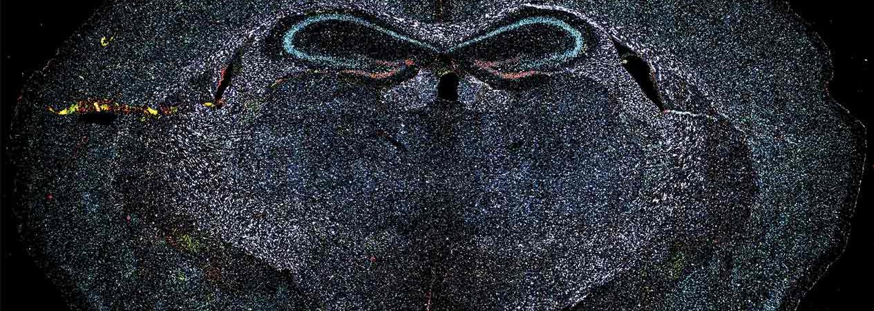
Spatial biology methods such as MERFISH, STARmap, seqFISH+ and CODEX rely on this evolution — and on laser sources that now reach into the near-infrared spectrum to expand the usable color palette without cross-talk between fluorophores.
Multicolor fluorescence imaging of mouse brain tissue showing spatial preservation of RNA species and nuclei. Image courtesy of Crest Optics (Italy) and Dr. Kwasi Kwakwa (Sanger Institute) >>

Capturing Biology as It Happens

Vesicle trafficking, calcium signaling, chromosome segregation: many biological processes unfold in milliseconds. Modern laser combiners enable rapid wavelength switching and precise intensity modulation, with diode lasers reaching rise and fall times of just a few tens of nanoseconds.
Synchronized with cameras and filter wheels, this level of control supports techniques like spinning disk confocal microscopy, letting researchers follow dynamic cellular events with minimal photobleaching.
<< Pulse-to-pulse repeatability of laser excitation measured at fiber output
Turning Imaging Into Reliable Data

As fluorescence microscopy is used more and more for quantitative measurement — comparing intensities across thousands of images or long time-lapse experiments — illumination stability becomes critical. Active optical feedback and precise power regulation now keep intensity fluctuations to a minimum over both short and long timescales, which is essential for high-content screening and extended live-imaging workflows in drug discovery, neuroscience and organoid research.
<< Normalized laser output power stability over 180 seconds, comparing active feedback control to a standard laser source
Matching Illumination to the Imaging Technique
Different modalities place different demands on laser light. Light-sheet microscopy benefits from stable alignment and efficient coupling for fast, low-phototoxicity imaging of large samples.
Laser spectrum before and after integration of a clean-up filter >>

TIRF microscopy requires excellent single-mode beam quality to generate the evanescent field used for studying membrane dynamics and single-molecule interactions. Advances in microstructured optical fibers now extend delivery from roughly 400–650 nm up to 405–1064 nm, opening the door to near-infrared fluorophores without redesigning the microscope.

Fiber ESM-5-125-PM developed and commercialized by Photonics Bretagne. A complete view of the fiber by SEM (left) and a zoom on the all-solid core surrounded by the air-holes microstructure (right). Courtesy of Photonics Bretagne, France
What’s Next: Adaptive and Large-Scale Illumination
Looking ahead, illumination is set to become smarter and more scalable: adaptive systems that adjust power, wavelength or timing in real time — increasingly guided by AI — to protect sample viability without missing key events; laser engines built to illuminate whole-organ and whole-brain samples evenly at higher power; and compact diode/DPSS modules bringing microscopy-grade stability into portable and clinical instruments, including fluorescence-guided surgery.
Laser illumination has quietly become one of the biggest levers for pushing fluorescence microscopy forward. Read the full article in BioPhotonics to see how these advances translate into practice.