
mesoSPIM Laser: How Oxxius Powers Tissue Clearing at INRAE’s IERP
Imaging an entire mouse brain, without sectioning it, down to the cellular level: that is what the first mesoSPIM microscope installed in France makes possible. The project was initiated by Christelle Langevin (IERP unit, INRAE), assembled by François Marquier and Karen Perronet’s team (ENS Paris-Saclay, LuMIn laboratory), and deployed within the IERP unit by Maxence Frétaud and Manon Mehraz, with an Oxxius laser at the heart of its optical train.
The challenge: imaging whole cleared samples, not slices
Biologists can now make entire organs, or even whole organisms, transparent using chemical tissue-clearing techniques. Once cleared, a sample can in theory be imaged in 3D by fluorescence, cell by cell, without sectioning. In theory only: conventional microscopes are designed for samples a few millimeters in size, not for a brain, a liver, or an entire mouse measuring several centimeters.
To overcome this bottleneck, the IERP (Experimental Infectiology of Rodents and Fish, an INRAE unit in Jouy-en-Josas led by Christelle Langevin) needed a light-sheet microscope capable of imaging large volumes at cellular resolution, without the cost or rigidity of commercial solutions. The answer came from a collaboration with physicists at the LuMIn laboratory (ENS Paris-Saclay / CNRS), built around mesoSPIM, an open-source microscope project.

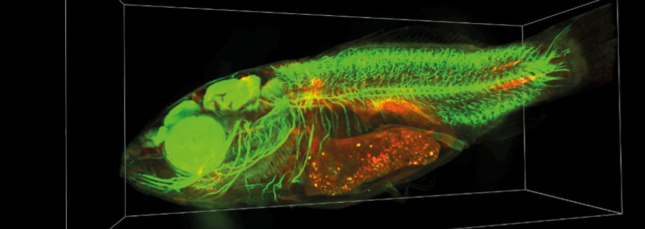
Cleared common carp labeled to reveal the nervous system (green). Autofluorescence is visible in red. Credits: IERP unit, INRAE.
The mesoSPIM: an open-source light-sheet microscope for large samples
The mesoSPIM (Mesoscale Selective Plane Illumination Microscope) is an instrument developed by an international scientific community, launched by Nikita Vladimirov’s team at the University of Zurich. Unlike turnkey commercial systems, it is built as a “kit” from freely shared plans and a parts list, which includes the Oxxius laser, explicitly recommended by the mesoSPIM initiative.
This approach is what convinced the IERP biologists (Christelle Langevin, Maxence Frétaud, Manon Mehraz) to take the plunge, working hand in hand with François Marquier, Karen Perronet, and the LuMIn team:
- Fast assembly: the microscope was built in two months, one to two days a week, by a second-year student from the Institut d’Optique.
- Controlled budget: up to 4 times more affordable than an equivalent commercial solution, thanks to kit-based assembly from open-source components.
- Benchtop configuration: a compact format designed from the outset to be portable and used for training.
- Cellular resolution: 1.5 µm lateral and 3.3 µm axial, on samples of several cubic centimeters.
Moved from ENS Paris-Saclay to the IERP in January 2025, six months ahead of schedule, the system was officially inaugurated on June 6, 2025. It is the first mesoSPIM installed in France.
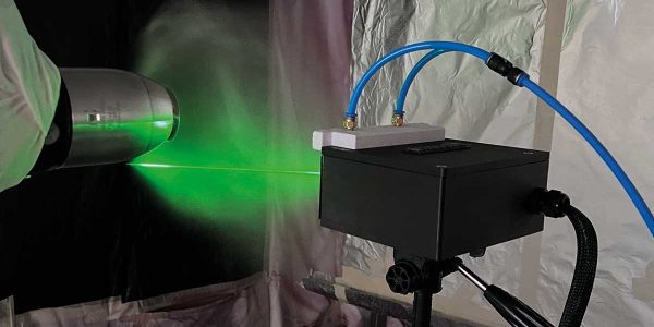
Picture on the right: The mesoSPIM Benchtop microscope, powered by an Oxxius laser combiner, installed at IERP-INRAE. Credits: IERP/ENS Paris-Saclay.

In their own words
The laser is fundamental to our optical setup. We wanted a robust system that was easy to integrate and offered the best value for money. When we saw that the mesoSPIM initiative recommended Oxxius, that immediately reassured us.
François Marquier, Professor at ENS Paris-Saclay, LuMIn laboratory.
Why an Oxxius laser?
In a light-sheet microscope like the mesoSPIM, the laser is the cornerstone of the optical train: it forms the light sheet, determines how uniform the illumination is, and ultimately governs the quality of the 3D reconstruction.
- Power stability: fluorescence intensity depends linearly on laser power. Over a volumetric acquisition lasting tens of minutes, even the slightest drift results in uneven brightness across the different planes of the sample, which is hard to correct afterward.
- Fiber-coupled output and multi-wavelength combining: several lasers are combined into a single fiber output, enabling fast switching between the colors needed for multicolor imaging.
- Adjustable power, tailored to each sample and each fluorescent marker.
- Recommended by the mesoSPIM initiative itself, through its open-source parts list: an immediate seal of confidence for the team.
- Rugged and reliable: the system has been transported by van many times.
- Responsive support: the Oxxius team stepped in directly during a training session to resolve a technical issue within minutes

The Oxxius laser combiner used on the IERP/ENS Paris-Saclay mesoSPIM: two outputs, two FlexxRay lasers (488 and 638 nm) and a StaxxBeam 561 nm single-frequency laser modulated by an AOM; total power: 350 mW. Credits: IERP/ENS Paris-Saclay.
Seamless integration with the mesoSPIM software
The Oxxius laser combiner is controlled directly by the mesoSPIM acquisition software, which manages wavelength changes throughout the acquisition. After a simple initial setup in the Oxxius software, laser control is fully integrated into the mesoSPIM software. No manual intervention is needed during imaging, so researchers can focus on their biological protocol rather than on the instrument.
The mesoSPIM by the numbers
1st
mesoSPIM microscope installed in France (inaugurated June 2025)
4x
cost up to 4 times lower than an equivalent commercial solution
1.5 / 3.3 µm
lateral / axial resolution, at the cellular scale on samples of several cm³
Tissue clearing applications in animal health
The mesoSPIM opens up new possibilities at the IERP that directly support its research missions in animal infectiology:
- Tracking the spread of a bacterial or viral infection throughout an entire organism.
- Visualizing the immune response, with the ability to quantify immune cells at the whole-organism level.
- Building volumetric datasets at cellular resolution, a particularly promising foundation for future artificial intelligence applications in computer-aided diagnosis, an area already attracting interest from AI researchers at CentraleSupélec.
Results recognized by the scientific community
Just four months after assembly began, the first biological samples were successfully imaged. The project has since gained visibility:
- La Recherche magazine published an article on the team’s work in tissue clearing, expansion microscopy, and light-sheet microscopy, led by Christelle Langevin (in French).
- The official inauguration established IERP/LuMIn as the first mesoSPIM site in France, among the roughly thirty systems listed worldwide at the time.The official inauguration established IERP/LuMIn as the first mesoSPIM site in France. As of September 1, 2026, more than 35 mesoSPIM systems were in operation worldwide (source: mesospim.org).
- The Benchtop system, transported several times for training sessions, has opened the instrument to the broader French scientific community beyond the founding team.
What’s next for the mesoSPIM: imaging even larger samples
The team is planning a mechanical upgrade of the mesoSPIM to accommodate even larger samples, on the order of 10 to 20 cm. This step up will likely come with new wavelengths, toward the red and infrared, to achieve deeper tissue penetration. In the longer term, coupling with spectrometry is being considered; still an exploratory avenue, it illustrates the mesoSPIM’s room for growth beyond its current uses. The team will also have to manage considerable data volumes: each volumetric acquisition at cellular resolution generates several terabytes of images.

Related products