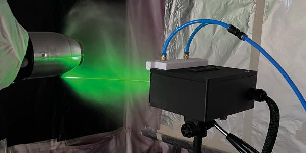
soSPIM Laser Combiner for Super-Resolution Microscopy: Single-Molecule 3D Imaging at IINS Bordeaux
Imaging individual fluorescent molecules inside intact living cells in 3D with a laser combiner for super-resolution microscopy that switches between two illumination modes in under one millisecond — that is the challenge Rémi Galland and his team at IINS Bordeaux tackle daily with the soSPIM platform, powered by an Oxxius L6Cc laser combiner.
What Is soSPIM and Why Does It Require a Multi-Wavelength Laser Combiner?

The soSPIM (single-objective Selective Plane Illumination Microscope) is a light-sheet microscopy platform developed by Rémi Galland during a post-doctoral fellowship between the Interdisciplinary Institute for Neuroscience (IINS, Bordeaux) and the MechanoBiology Institute (Singapore). It was patented and published in Nature Methods in 2015 (Galland et al., Nature Methods, 2015, vol. 12(7), pp. 641–644).
Unlike conventional light sheet microscopy systems that require two opposing objective lenses, soSPIM uses a single objective lens combined with microfabricated devices embedding arrays of micro-wells flanked with micromirrors. These micromirrors redirect the excitation beam perpendicular to the optical axis, creating a light sheet inside the sample without a second objective: a geometry that makes soSPIM fully compatible with standard inverted microscopes, while opening up a wide range of sample formats, from single suspended cells to whole organisms.
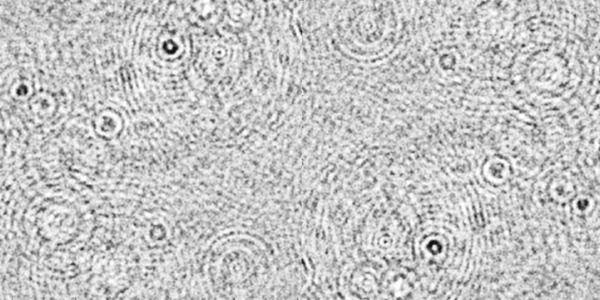
soSPIM multi-color single-molecule super-resolved image of the nucleus envelope (LaminB1) and Golgi apparatus of a suspended T cell. Multi-color soSPIM super-resolution image acquired using the Oxxius L6Cc laser combiner. Credit: R. Galland, C. Butler, JB. Sibarita – Univ. Bordeaux / CNRS / IINS
This single-objective architecture also imposes specific requirements on the laser source: the illumination must be both powerful enough to image deep into the sample and stable enough to sustain quantitative measurements over hours to days. And because soSPIM is combined with TIRF on the same microscope body, the laser source must switch instantaneously between two independent output paths, without any manual intervention.
soSPIM imaging capabilities at a glance
- Single-molecule sensitivity down to the individual fluorescent protein level
- 3D imaging depth from the coverslip surface up to tens of microns inside the sample
- Sample format flexibility: single suspended cells, cell doublets, 3D aggregates, whole organisms
- Two combined modalities on one microscope: soSPIM light-sheet + TIRF illumination
- Temporal range: from millisecond dynamics to multi-day organoid growth monitoring
This dual-modality design is precisely why a high-performance, multi-wavelength laser combiner with fast output switching is required.
The Scientific Context: Studying Synapse Transmission at the Molecular Scale
The Interdisciplinary Institute for Neuroscience (IINS) is part of Bordeaux Neurocampus, a European centre of excellence in neuroscience. IINS brings together biologists, neurobiologists, chemists, biophysicists, and optical scientists to study synapse transmission from the molecular scale to the whole organism.
Within IINS, the Quantitative Imaging of the Cell team led by Jean-Baptiste Sibarita develops cutting-edge quantitative imaging methods to decipher protein organization and dynamics with high spatial and temporal resolution — and transfers these methods, both academically and industrially, to the broader scientific community.
In their own words
In our team we typically represent the interdisciplinary aspect of IINS. Our main goal is to develop cutting-edge quantitative imaging methods combining optics, image analysis and bioengineering to decipher protein organization and dynamics at the nanoscale.
Rémi Galland, CNRS Researcher, Quantitative Imaging of the Cell, IINS Bordeaux Neurocampus
Why Choose the Oxxius L6Cc Laser Combiner for Super-Resolution Microscopy?
The soSPIM platform operates in two distinct illumination modes: soSPIM light-sheet imaging for in-depth 3D acquisition, and TIRF illumination for single-molecule super-resolution imaging at the coverslip surface. Both modes share the same microscope body and must switch seamlessly during an experiment.
This dual requirement drives three non-negotiable laser source criteria:

1. Multi-wavelength integration in a single compact housing
The soSPIM requires simultaneous access to 405 nm, 488 nm, 561 nm and 640 nm laser lines to cover standard fluorescent protein and dye combinations used in super-resolution microscopy (SMLM, STORM, PALM).
2. Fast, automated switching between two fiber outputs
Switching between soSPIM and TIRF optical paths must occur without manual intervention, in under 1 ms, at rates up to 30 Hz, synchronized with camera exposure.
3. Long-term power stability across extended acquisition sessions
Organoid growth monitoring runs for hours to days. Any drift in laser power translates directly into quantification errors. A stability of ±2 % over 8 hours is the accepted threshold.
The Oxxius L6Cc laser combiner paired with the MDL-FSTM fast-switch mirror extension module meets all three criteria in a single, fiber-coupled system.
Oxxius L6Cc Configuration at IINS Bordeaux
Oxxius Laser combiner: MixxWave L6Cc
Ultra-compact, all-in-one multi-wavelength combiner (analog and TTL modulation) compatible with µManager, LabVIEW and MetaMorph.
Laser lines integrated at IINS:
| Wavelength | Technology | Power | Options |
|---|---|---|---|
| 405 nm | Laser diode | 50 mW | — |
| 488 nm | Laser diode | 100 mW | Clean-up filter |
| 561 nm | DPSS | 150 mW | Electromechanical shutter + AOM |
| 640 nm | DPSS | 500 mW | Electromechanical shutter |
Fast-switching module: MDL-FSTM-L6Cc
| Specification | Value |
|---|---|
| Max switching rate | 30 Hz |
| Switching time | < 1 ms |
| Wavelength range | 375–785 nm |
Dual fiber output
| Specification | Value |
|---|---|
| Coupling efficiency | > 70% |
| Fiber type | Polarization-maintaining (RGBV) |
| Connector | FC/APC |
| Power stability (8 h) | ±2% |
The L6Cc delivers its combined beam via a polarization-maintaining fiber-coupled output (FC/APC connector, coupling efficiency > 70%, RGBV). This fiber-coupled laser architecture is a critical integration advantage: it forms the foundational starting point of the soSPIM optical path, ensuring spatial mode quality and mechanical decoupling between the laser source and the microscope body.
In their own words
The soSPIM technology requires both powerful and very stable laser lines over time, at multiple wavelengths. We also needed to combine two imaging modalities (soSPIM and TIRF) on the same microscope. The Oxxius L6Cc with the MDL-FSTM extension module was the right answer: it lets us switch instantly between our two illumination paths, with the stability we need for single-molecule imaging sessions.
Rémi Galland, CNRS Researcher, Quantitative Imaging of the Cell, IINS Bordeaux Neurocampus
Applications: From Single-Molecule Super-Resolution to Organoid Screening
Super-resolution imaging at the single-molecule level
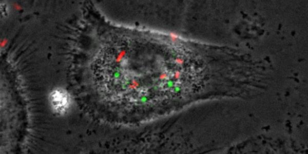
The soSPIM enables multi-color single-molecule localization microscopy (SMLM) with nanometric resolution inside intact cells. Rémi Galland’s team has demonstrated the imaging of complete subcellular structures — the full nuclear envelope (LaminB1) and Golgi apparatus of suspended T cells — with near-molecular precision in three dimensions, tens of microns above the coverslip.
3D live imaging and organoid growth monitoring

Combined with newly developed microfabricated culture devices, the soSPIM monitors the growth of stem cell cysts and organoids in real time, in 3D, over multiple days. The system is currently being developed into a high-content screening platform to standardize and parallelize the culture and imaging of organoids — unlocking their full predictive potential in pharmacology and disease modelling.
3D soSPIM image of a stem cell cyst grown in a capsule (TreeFrog Therapeutics© technology). Color-coded by imaging depth.
Credit: R. Galland, C. Butler, JB. Sibarita – Univ. Bordeaux / CNRS / IINS.
Key Results Enabled by the Oxxius L6Cc Laser Combiner
<1 ms
Switching time between soSPIM and TIRF outputs
Instant, automated switching – no manual realignment
±2 %
Laser power stability over 8 hrs
The decisive criterion for long-term single-molecule acquisitions
4
Wavelengths in a single compact combiner
Simultaneous laser lines covering all fluorophores used in cell biology
<1 ms switching time between soSPIM and TIRF modes: enabling seamless multiplexed acquisition protocols on the same microscope without any manual realignment.
Single-molecule sensitivity: the power and stability of the four integrated laser lines enable detection of individual fluorescent molecules tens of microns inside living cells.
±2 % power stability over 8 hours: sustaining consistent illumination across multi-hour organoid growth monitoring sessions, where photon budget consistency is critical for quantitative analysis.
4 simultaneous wavelengths (405 nm / 488 nm / 561 nm / 640 nm) in a single compact housing: providing full spectral coverage for multi-color super-resolution and live-cell imaging.
Outlook
The next development phase of the soSPIM platform focuses on engineering a fully parallelized, high-content organoid imaging system, combining standardized microfabricated culture devices with the soSPIM’s 3D live-imaging capabilities to quantify disease phenotypes and drug responses across large organoid populations.
The Oxxius MixxWaver L6Cc laser combiner, with its multi-wavelength integration, sub-millisecond output switching and long-term power stability, remains the laser source of choice as the platform scales toward broader scientific and translational applications in neuroscience and drug discovery.