Interview with IINS Bordeaux neurocampus
Interview with Rémi Galland, Researcher CNRS
The soSPIM technology
[Interview with]
Rémi Galland, Researcher CNRS
Hi Rémi, could you introduce yourself and give us a quick understanding of what IINS Bordeaux Neurocampus is ?
Hi Oxxius, I am 39 years old and I am a researcher in optics at the Interdisciplinary Institute for Neuroscience in Bordeaux headed by Daniel Choquet in the team Quantitative Imaging of the Cell directed by Jean-Baptiste Sibarita.
IINS is part of the Bordeaux Neurocampus which is a European centre of excellence on neuroscience gathering pluridisciplinary research teams studying brain functions and its pathologies from the molecular scale up to the whole animal or human brain scale. At IINS we are more precisely studying synapse transmission at the molecular and cellular scale using interdisciplinary approaches associating biologists, neurobiologists, chemists, biophysicists and opticians.
I like to say that in the team Quantitative Imaging of the Cell we typically represent this interdisciplinary aspect of IINS. Indeed, our main goal is to develop cutting-edge quantitative imaging methods combining optics, dedicated image analysis methods and bioengineering techniques to decipher protein organization and dynamics with high spatial and temporal resolution. Those methods are then applied to address specific biological questions in close collaboration with biology research groups. We also have an important academic and industrial technological transfer activity. This is typically the case with the soSPIM technology we recently developed and would like to share with the scientific community.
Could you explain in few words what the soSPIM technology is ?
The soSPIM technology, for single-objective Selective Plane Illumination Microscope, is a unique system allowing 3D imaging of biological samples at high spatial and super resolution and various temporal scales. It arises from a collaboration in between our team at IINS and the team Biomechanics of Cell-cell Contacts headed by Virgile Viasnoff at the MechanoBiology Institute in Singapore. In 2013 and 2014, I performed a Post-doc in between this two teams which enabled me to develop and demonstrate the imaging capacities of the soSPIM system we patented and published in 2015 in the journal Nature Methods (Galland et al., Nature Methods, 2015, vol. 12(7), pp 641-644).
This technology relies on microfabricated devices embedding arrays of micro-wells flanked with micromirrors that enable the creation of a light-sheet perpendicular to the objective optical axis. We demonstrated its capability to image biological samples of various dimensions (from single cell, cell doublet, 3D cell aggregates up to whole organisms) with sensitivity down to the single molecule level.
Combining the soSPIM technology with newly developed microfabricated devices, we are now engineering a versatile high-content screening platform to standardize and parallelize both the culture and the 3D live imaging of the emerging organotypic 3D cellular cultures (ie Organoids). Such a platform will enable to quantify disease and drug induced variability in organotypic cultures, exploiting organoid full predictive potential in health science.
You choose to use the Oxxius L6Cc with the extension module MDL-FSTM. Can you explain why and share your feedback ?
The soSPIM technology enables to monitor biological events at various temporal scale up to days but also to perform single molecules based super-resolution imaging tens of microns above the coverslips to image whole cells with near molecular resolution. Therefore, it requires both powerful and very stable laser lines along time at various wavelength. In addition, we choose to combine on a same microscope the soSPIM technology with a TIRF illumination system in order to combine standard and in-depth single molecule based super-resolution imaging. That’s why we choose a L6Cc combiner from Oxxius with the extension module MDL-FSTM which enables to easily switch in between our two imaging modalities.
ZOOM ON
|
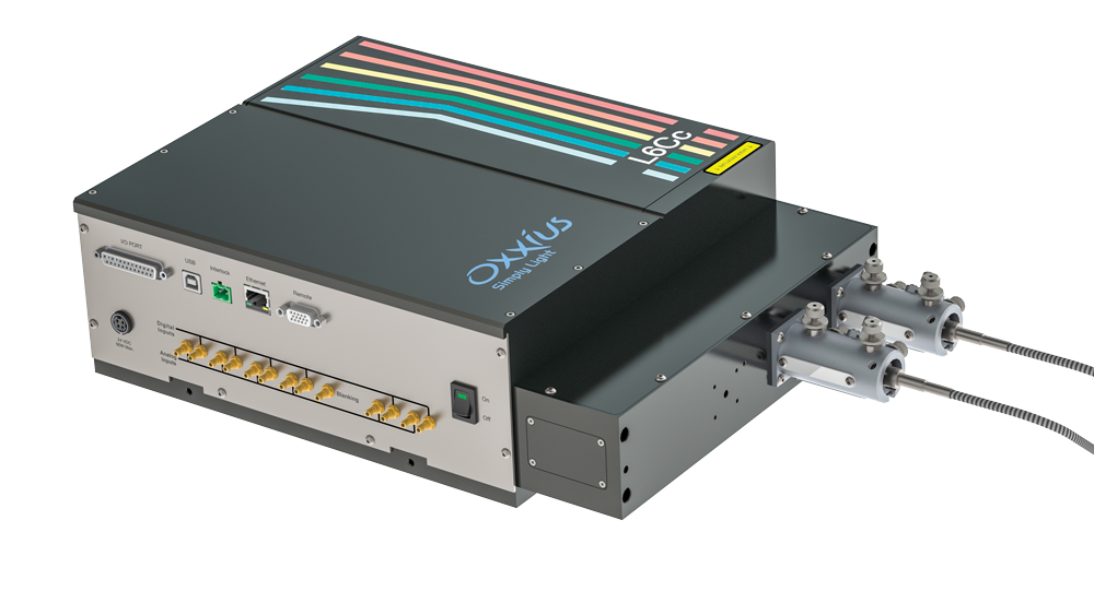 |
All-In-One combinerFlexible and upgradeable – Analog and TTL modulation – Compact, power-efficient system – Compatible µmanager, Labview, Metamorph
|
IINS configuration:– Laser diode 405 nm, 50 mW – Laser diode 488 nm, 100 mW, Clean up filter 488 nm – Laser DPSS 561 nm, 150 mW with Electromechanical shutter and AOM – Laser DPSS 640 nm, 500 mW with Electromechanical shutter
|
MDL-FSTM-LnCc,
|
Dual Output fiber– Coupling efficiency >70% – Polarization maintaining fiber RGBV – Connector FC/APC – Power stability (over 8 hours) ±2% |
What is the field of applications ?
Do you already have some pictures to communicate ?
The soSPIM technology has a wide range of applications from in-depth single-molecule based super-resolution imaging up to the monitoring of the growth of complex 3D cellular cultures. Here I would like to share two pictures illustrating these two highly different imaging capacities:
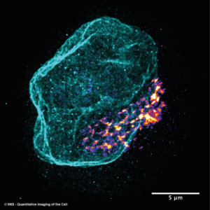
soSPIM Multi-color single molecule based super-resolved image of the whole nucleus envelop (LaminB1) and Golgi apparatus of a suspended T cell.
Credit : Image courtesy of R. Galland, C. Butler and JB. Sibarita – Univ. Bordeaux, CNRS, Interdisciplinary Institute for Neuroscience, IINS – Image acquired using the soSPIM technology
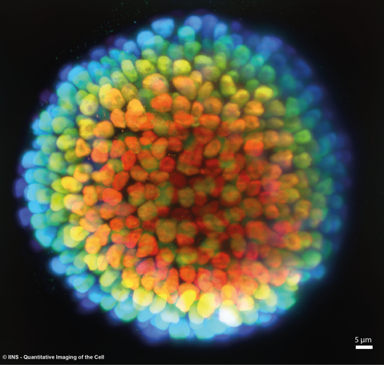
3D soSPIM picture of a stem cells cyst grown in a capsule using the TreeFrog Therapeutics© Technology. The color code for the imaging depth. Credit : Image courtesy of R. Galland, C. Butler and JB. Sibarita – Univ. Bordeaux, CNRS, Interdisciplinary Institute for Neuroscience, IINS – Image acquired using the soSPIM technology
 |
 |